



 切换语言
切换语言
切换语言
切换语言
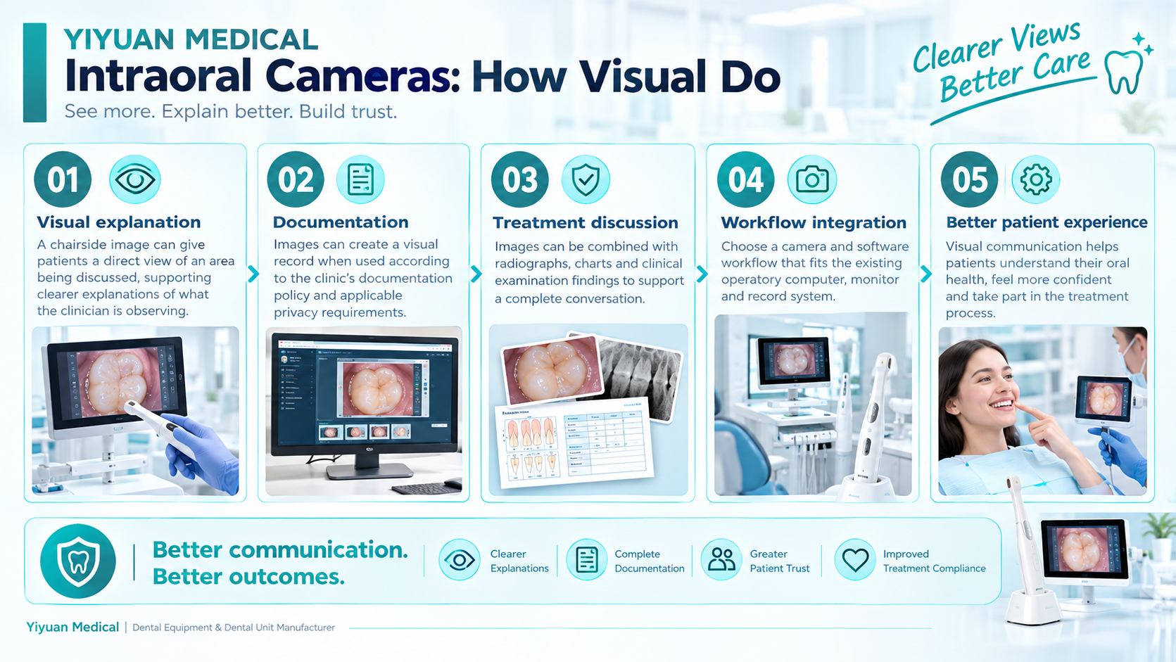
Dental decisions often involve information that is difficult for patients to see directly. Intraoral cameras can add a visual layer to the clinical conversation. They do not replace clinical examination or other diagnostic tools, but they can make it easier to show the exact area being discussed. This is especially relevant in a world where oral disease remains a large public-health burden: WHO estimates that oral diseases affect nearly 3.7 billion people globally.
A patient may understand a verbal explanation differently after seeing an enlarged image of the relevant tooth or soft-tissue area. A camera can make the conversation more concrete by linking the clinician’s explanation to a visible location. The benefit is communicative; the device should not be marketed as independently diagnosing conditions unless the specific device has the regulatory authorization and clinical evidence to support that claim.
The U.S. CDC reports that about 21% of adults aged 20–64 had at least one untreated cavity in the 2017–March 2020 surveillance period. NIDCR likewise reports that nearly 90% of U.S. adults aged 20–64 had experienced dental decay. These data illustrate the scale of unmet or historical oral-health needs, but they do not prove that intraoral cameras reduce disease rates. The appropriate message is that visual tools can support communication and documentation within a professional clinical workflow.
Look at image quality, focus range, lighting, ergonomics, cable or wireless configuration, disinfection compatibility, software connectivity, image export options and integration with the practice-management or imaging workflow. Ask how the device is cleaned between patients and what parts are replaceable.
A practical workflow is: capture -> review -> annotate when appropriate -> explain -> document -> store or export according to the clinic’s policy. This keeps the camera from becoming a standalone gadget and makes it part of a repeatable patient-engagement process.
Patient images are health information in many jurisdictions. Clinics should follow the applicable privacy law, consent policies and record-retention rules for their location. Product literature should avoid suggesting that image capture alone meets every privacy or documentation requirement.
What is an intraoral camera used for?
It can provide enlarged visual images that support patient explanations and documentation within the clinic workflow.
Can an intraoral camera replace diagnosis?
No. Its role and claims depend on the device and regulatory status; visual capture should complement professional clinical assessment.
Evidence & source note
|
Organization / evidence |
Original source address |
|
World Health Organization (WHO) |
https://www.who.int/news-room/fact-sheets/detail/oral-health |
|
U.S. NIDCR |
https://www.nidcr.nih.gov/research/data-statistics/dental-caries/adults |
|
U.S. CDC |
https://www.cdc.gov/oral-health/php/2024-oral-health-surveillance-report/selected-findings.html |